
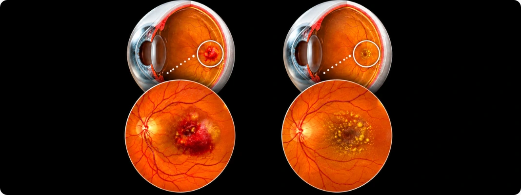
According to the American Academy of Ophthalmology, about 80% of people with age-related macular degeneration have the dry form, making it much more common than wet AMD. AMD is also a leading cause of vision loss in people aged 50 and older, although some people may have early changes before noticing a problem with their sight.
You may notice that reading takes more effort, straight lines look different, or the center of your vision is not as sharp as it used to be, even when your contact lenses otherwise provide clear refractive correction. With wet vs dry macular degeneration, knowing how the two forms differ can help you recognize changes that need an eye doctor rather than trying to diagnose the problem yourself.
Quick Summary
- Dry AMD is the more common form and usually progresses gradually, while wet AMD can cause faster changes in central vision.
- Both types affect the macula and can make reading, recognizing faces, driving, and seeing fine details more difficult.
- Dry AMD may progress to geographic atrophy or, in some people, convert to wet AMD.
- Macular degeneration treatment depends on the type and stage and may include monitoring, AREDS2 supplements for eligible patients, geographic atrophy treatment, or anti-VEGF injections for wet AMD.
- New wavy lines, a central dark or blank spot, or sudden worsening of vision should be evaluated promptly by an eye care professional.
What is the difference between wet and dry macular degeneration?
Dry AMD usually develops gradually as drusen, pigment changes, and thinning or loss of cells in the macula affect the retina. Wet AMD develops when abnormal blood vessels grow beneath or within the retina and leak fluid or blood, which can damage the macula and cause faster central vision loss. Advanced dry AMD can also cause substantial vision loss through geographic atrophy, while wet AMD is considered an advanced form of the disease.
The main difference between wet and dry macular degeneration is therefore not simply which one is “worse.” They differ in what is happening inside the retina, how quickly vision may change, and which treatment options may be appropriate, differences that vision correcting contacts cannot address because AMD involves the retina.
Symptoms can overlap, so only a comprehensive eye examination and retinal imaging can determine which type you have.
| FEATURE | DRY AMD | WET AMD |
|---|---|---|
| What Happens | Drusen, pigment changes, and macular thinning or atrophy | Abnormal blood vessels grow and leak fluid or blood |
| How Common | More common, about 80% of AMD cases | Less common |
| Typical Pace | Often gradual | Can progress rapidly |
| Early Symptoms | Often none | Changes may appear more suddenly |
| Vision Affected | Central vision | Central vision |
| Advanced Form | Geographic atrophy | Neovascular or exudative AMD |
| Main Treatment Approach | Monitoring, AREDS2 for eligible patients, and treatment for selected cases of geographic atrophy | Anti-VEGF injections and, in selected cases, other therapies |
| Urgency | Regular monitoring and prompt care for new changes | Prompt evaluation and treatment |
Note: These are typical patterns, not a way to diagnose AMD yourself. Advanced dry macular degeneration can cause serious central vision loss, and symptoms of wet and dry macular degeneration can overlap, so new or worsening vision changes should be evaluated by an eye care professional.
What does macular degeneration do to your central vision?
Age-related macular degeneration damages the macula, the small central part of the retina that helps you see fine details directly in front of you. As the macula changes, you may develop blurry, distorted, or missing areas in your central vision, making it harder to read, recognize faces, drive, or see detailed objects. Your side, or peripheral, vision is generally preserved, so AMD does not typically cause complete blindness.
That distinction matters because AMD-related blur is not the same as an ordinary focusing problem. A refractive error vs visual impairment such as nearsightedness, farsightedness, or astigmatism can often be sharpened with glasses or contact lenses, while AMD involves damage to retinal tissue itself. Changing your prescription may help with a separate refractive error, but it cannot correct central vision loss caused by macular degeneration.
Vision changes also vary from person to person. Some people with early AMD may notice little or no change for years, while others develop more noticeable difficulty with detailed straight-ahead vision as the condition progresses. Regular eye exams matter because AMD can be present before you notice obvious symptoms.
What do wet and dry macular degeneration symptoms look like?
Wet and dry macular degeneration symptoms can overlap, especially once AMD reaches a later stage, so you cannot reliably tell which type you have from symptoms alone. Early dry AMD often causes no symptoms, while intermediate disease may bring mild central blur or difficulty seeing in low light. With late dry or wet AMD, straight lines may look wavy or crooked, colors can appear less bright, and blurry or blank areas may develop near the center of your vision.
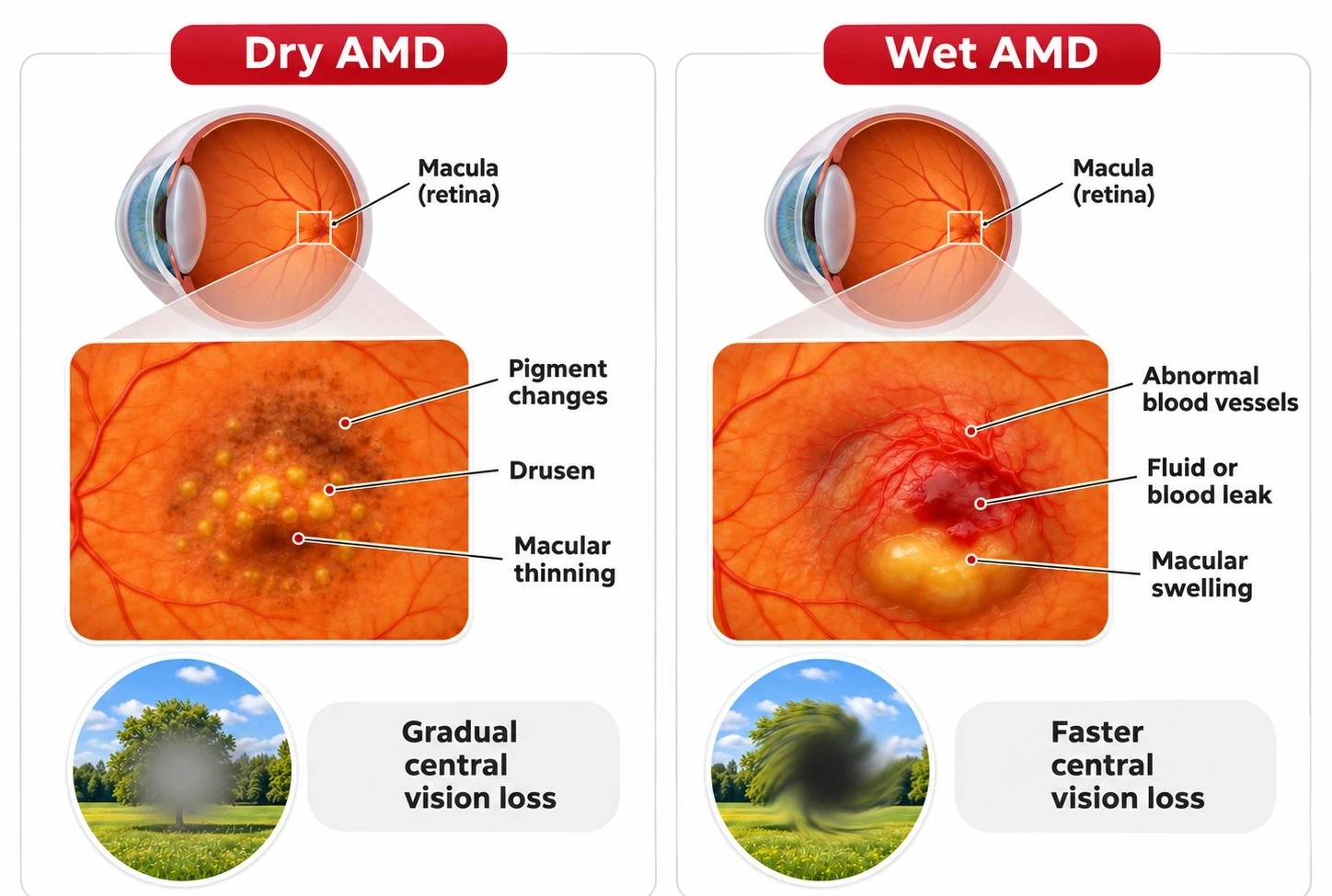
The main difference is often the pace of change. Dry AMD generally progresses more gradually, while wet AMD can cause faster vision changes because abnormal blood vessels grow beneath the retina and may leak fluid or blood. Any new distortion or sudden change in your central vision deserves prompt evaluation by an eye doctor rather than assuming your Rx contacts need changing.
What can dry macular degeneration feel like?
Dry macular degeneration symptoms depend on the stage. Early dry AMD may feel completely normal, which is why it can first be detected during an eye exam rather than because you noticed a problem. Intermediate dry AMD may cause subtle blurring in the center of your vision or make dim lighting more challenging.
As dry AMD advances, changes can become more noticeable. Geographic atrophy, an advanced form of dry AMD in which areas of retinal cells are lost, can create increasingly impaired or missing areas in your central vision.
You may notice:
- Mild or increasing blurry central vision
- More difficulty reading or seeing fine details
- Greater difficulty seeing in dim lighting
- Colors that seem less bright than before
- Wavy or crooked-looking lines in later disease
- Blurry or blank areas near the center of your vision
Finding low-light tasks harder than they used to be? Explore why driving at night can become more difficult to understand other factors that can affect vision after dark.
What can wet macular degeneration feel like?
Wet macular degeneration symptoms often change more quickly than those of dry AMD, although not every case begins suddenly. Because abnormal blood vessels can leak beneath the retina, you may notice that lines or edges that should be straight look bent or wavy, or that a new blurry or blank area appears in your central vision. Reading, recognizing faces, and seeing fine details may also become noticeably harder over a shorter period.
You may notice:
- Newly wavy, bent, or crooked straight lines
- A new blurry area in the center of your vision
- Blank or missing spots in central vision
- A noticeable decline in detailed vision
- Colors that look less bright
- Greater difficulty reading or recognizing faces
These symptoms do not prove that you have wet AMD because other eye conditions can also change central vision. If straight lines suddenly look wavy, a new central spot appears, or your vision worsens noticeably, contact an eye care professional promptly rather than waiting for your next routine exam.
How fast can wet and dry macular degeneration progress?
Dry AMD progression is usually gradual, but there is no fixed timeline because the condition can change differently from one person to another. Some people stay in the early or intermediate stages for a long time, while others progress to geographic atrophy, an advanced form of dry AMD in which areas of retinal cells stop functioning and central vision can become more impaired. Dry AMD can also turn into wet AMD, although this does not happen to everyone.
Wet AMD can affect vision much faster because abnormal blood vessels grow beneath the retina and may leak fluid or blood into the macula. You may notice new blur, distortion, or straight lines that suddenly look wavy or bent. Because these changes can happen quickly, contact an eye doctor promptly if you notice a new or sudden change in your central vision, even if your eyesight had seemed stable with prescription contact lenses before.
What raises your risk of macular degeneration as your eyes age?
Increasing age is the strongest risk factor for age-related macular degeneration, but it is not the only one. Smoking and family history are well-established risks, and conditions linked with cardiovascular health, including high blood pressure and high cholesterol, are also associated with AMD.
Some risk factors are outside your control, while others give you practical areas to discuss with your healthcare team.
Factors you cannot change include:
- Getting older
- Having a family history of AMD
- Genetic susceptibility
- Some demographic risk differences identified in population studies
Factors you may be able to address include:
- Smoking or exposure to tobacco
- High blood pressure
- High cholesterol
- Excess body weight
- A diet high in saturated fat
- Low levels of regular physical activity
If you have several risk factors, ask your eye care professional how often you should have comprehensive eye exams. Quitting smoking, staying physically active, eating a balanced diet, and managing blood pressure and cholesterol with your healthcare team may help lower risk, but none of these measures should be described as a cure or guaranteed prevention for AMD.
Not sure how often your eyes should be checked as you get older or accumulate AMD risk factors? Review how often you need a comprehensive eye exam and follow the schedule your eye care professional recommends for your individual risk.
How does an eye doctor tell wet AMD from dry AMD?
An eye doctor cannot reliably distinguish wet from dry AMD from symptoms alone or from how clearly you see with prescription contact lenses. A dilated retinal examination and optical coherence tomography OCT imaging allow your ophthalmologist to examine the macula for findings such as drusen, pigment changes, retinal thinning or atrophy, fluid, and other structural changes. BMJ Best Practice identifies OCT as the most widely used imaging method for diagnosing and managing AMD.
Your evaluation may include:
- A dilated retinal exam so the doctor can inspect the retina and macula for drusen, pigment changes, bleeding, fluid, or atrophy. If you are unfamiliar with dilation, see what to expect when getting your eyes dilated.
- Optical coherence tomography or OCT, a noninvasive scan that uses light waves to create detailed cross-sectional images of the retina. It can help show retinal thinning, fluid, swelling, and other changes associated with AMD.
- OCT angiography or OCTA, which creates images of blood vessels in and beneath the retina without injecting dye. It can help your doctor examine abnormal vessel growth associated with wet AMD.
- Fluorescein angiography when a closer look at retinal circulation or leaking abnormal blood vessels is needed. A dye is injected into a vein while specialized retinal photographs track how it moves through the eye’s blood vessels.
- An Amsler grid to identify central distortion, blurred areas, or missing spots that need further evaluation. The grid can help you and your doctor monitor changes, but it cannot diagnose wet or dry AMD by itself.
Because AMD requires examination of the retina, an online vision check is not a substitute for a comprehensive retinal evaluation when macular disease is suspected.
Considering an online vision test because it is more convenient? Check what states allow online eye exams while remembering that suspected retinal disease still requires an in-person evaluation.
How are wet and dry macular degeneration treated differently?
Macular degeneration treatment depends on the type and stage of AMD, while vision correcting contacts address separate refractive needs rather than the retinal disease itself. Early and intermediate dry AMD are generally managed with monitoring and, for certain patients, AREDS2 supplements, while advanced dry AMD with geographic atrophy now has injectable treatments that can slow the growth of atrophic areas. Wet AMD is treated more urgently, most often with anti-VEGF injections that target the abnormal blood-vessel growth and leakage responsible for rapid vision changes.
How is early or intermediate dry AMD managed?
Early dry AMD is usually monitored rather than treated with medication. Your eye care professional may recommend regular dilated eye exams, healthy lifestyle habits, and home vision checks to watch for changes. AREDS2 supplements for AMD are not meant to prevent AMD or treat everyone with early disease, but they may help certain people with intermediate AMD or advanced AMD in one eye.
In the high-risk groups studied by the National Eye Institute, the AREDS and AREDS2 formulations reduced the risk of progressing to advanced AMD by about 25%. This benefit applies to a specific combination of nutrients and specific stages of AMD, not to ordinary multivitamins or any supplement marketed for “eye health.” Ask your eye doctor whether AREDS2 is appropriate for you before starting high-dose supplements.
What treatments are available for geographic atrophy?
Geographic atrophy treatment has changed in recent years. Two FDA-approved complement-inhibiting medicines, Pegcetacoplan (Syfovre) and Avacincaptad pegol (Izervay), can be injected into the eye to slow the enlargement of geographic atrophy, an advanced form of dry AMD. These treatments slow disease progression but do not regrow lost retinal cells or restore vision that has already been lost.
Treatment also involves a real tradeoff between potential benefit and the burden and risks of repeated eye injections. Your retina specialist will consider factors such as the location and size of the atrophy, how quickly it is progressing, your remaining vision, and possible treatment complications before recommending injections. These medicines are a treatment option, not a cure.
How is wet AMD treated?
The main wet macular degeneration treatment is anti-VEGF therapy, which uses injections into the eye to slow the growth and leakage of abnormal blood vessels beneath the retina. These medicines block VEGF, a protein that contributes to abnormal blood-vessel growth in wet AMD. Treatment usually requires repeated injections and retinal monitoring, with the schedule adjusted based on the medicine used and how your eye responds.
Anti-VEGF treatment can help stabilize vision, and some people may regain some vision, especially when treatment starts before extensive retinal damage or scarring develops. However, it cannot guarantee that all lost vision will return, so you should not delay or stop treatment simply because your vision seems stable. Photodynamic therapy is still used in selected cases, sometimes alongside anti-VEGF injections, but it is much less common today.
How can you monitor AMD between eye appointments?
An Amsler grid is a simple home-monitoring tool that can help you notice new changes in your central vision between appointments. It may reveal areas where straight lines suddenly look wavy, blurred, dark, or missing, but it cannot diagnose dry or wet AMD on its own. Many ophthalmologists recommend checking daily when you have AMD, although you should follow the schedule your own eye care professional gives you.
- In good lighting, put on the glasses or other near correction you normally use for reading and hold the grid at your usual reading distance, roughly 12 to 15 inches away.
- Cover one eye and look directly at the center dot with the uncovered eye. Keep your gaze fixed on that point rather than scanning around the grid.
- While looking at the center, notice whether any lines appear newly wavy, bent, blurry, dark, blank, or missing. Then repeat the same check with your other eye.
- If you notice a new or worsening change, contact your ophthalmologist promptly. Do not wait for your next routine appointment to report a new central dark spot, distortion, or missing area.
The Amsler grid is useful for home monitoring, but it does not replace dilated retinal exams, optical coherence tomography OCT, or scheduled treatment visits.
Can contact lenses help if you have macular degeneration?
Contact lenses cannot treat macular degeneration or restore central vision loss caused by retinal damage. They may still correct a separate refractive error with contacts for myopia, contact lenses for farsightedness, or another prescribed option if your eye care professional determines that contacts are appropriate for you. However, vision impairment from AMD may remain even with the best glasses or contact lens prescription because the problem involves the retina rather than the eye’s ability to focus light. The National Eye Institute defines low vision as vision loss that can continue even with glasses or contact lenses.
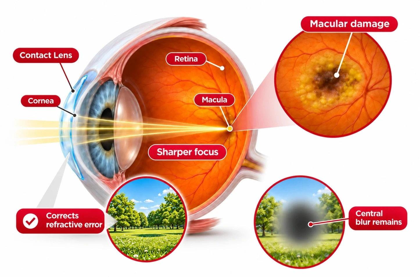
That difference is why a changing prescription does not always explain a new blur. If you want a closer look at how these conditions differ, Lens.com explains refractive error vs visual impairment and why some types of vision loss cannot be corrected with lenses alone.
If you wear contacts and suddenly notice a new central blind spot, wavy straight lines, or an unexplained change in one eye, do not assume the lens is simply dirty or that your prescription has changed. Remove your contacts, switch to backup glasses if you have them, and seek prompt professional evaluation if the abnormal vision remains. Sudden vision changes can have several causes and need an eye examination rather than repeated contact lens changes.
Still need contact lenses for a separate refractive error while managing AMD? Know how to read contact lens prescriptions (OD vs. OS) so the prescription values for your right and left eyes stay correctly matched.
When should you seek prompt or emergency eye care?
Contact an ophthalmologist or retina specialist promptly if you develop a new or noticeably worsening change in your central vision. New distortion can occur with wet AMD, but it can also have other causes, so symptoms alone cannot tell you what is happening inside the eye. Do not wait for your next routine appointment or assume your prescription contact lenses need changing if the vision change is sudden or clearly different from your usual vision.
Warning signs that need prompt professional evaluation include:
- Newly wavy, bent, or distorted straight lines
- A new dark, gray, blurry, or blank area in your central vision
- Rapidly worsening central blur
- A noticeable sudden change in vision in one eye
- A sudden decline in reading or detailed vision
- New distortion that develops when you already have dry AMD
A sudden major loss of vision is an emergency, even if you do not know the cause. Seek emergency medical care immediately, especially if the vision change occurs with facial drooping, weakness or numbness on one side, trouble speaking or understanding speech, loss of balance, or a sudden severe headache, because these can be signs of stroke; in the U.S., call 911 rather than driving yourself to the hospital.
Unsure whether a new vision change can wait for your next routine appointment? Review common reasons to see an eye doctor so you can recognize symptoms that deserve earlier professional evaluation.
What should you know about wet vs dry macular degeneration?
Dry AMD is more common and usually changes vision more gradually, while wet AMD can cause faster central vision loss when abnormal blood vessels grow and leak beneath the retina. Both forms can interfere with reading, recognizing faces, driving, and other tasks that depend on detailed central vision, and an eye examination with retinal imaging is needed to determine the type and stage. Macular degeneration treatment may include monitoring, AREDS2 supplements for appropriate patients, injections that slow geographic atrophy in selected patients, or anti-VEGF therapy for wet AMD. New wavy lines, a central dark or blank spot, or a sudden change in vision should be evaluated promptly rather than saved for your next routine appointment.
Do you also need vision correction for nearsightedness, farsightedness, astigmatism, or presbyopia while managing your eye health? Browse our contact lenses to compare prescription options for your refractive needs, keeping in mind that contact lenses do not treat macular degeneration.
Learn More About Wet and Dry Macular Degeneration
Get clear answers to common questions about wet and dry macular degeneration, including which type can progress faster, whether vision loss can be reversed, how dry AMD can change over time, and when you may need closer eye monitoring.
Which is worse to have dry or wet macular degeneration?
Wet AMD is generally more urgent because it can cause central vision loss more quickly, but advanced dry AMD can also cause severe permanent visual impairment. Wet AMD involves abnormal blood vessels that can leak fluid or blood beneath the retina, while advanced dry AMD can lead to geographic atrophy and loss of retinal cells. Calling one type simply “worse” can therefore be misleading because the speed, stage, and amount of retinal damage all matter. Both forms require appropriate monitoring, and new vision changes should be reported promptly.
Can dry macular degeneration be fixed?
No, dry AMD cannot currently be reversed. Early disease is generally monitored, and AREDS2 supplements for AMD may slow progression in people with certain intermediate or advanced stages, but they do not benefit everyone and cannot prevent AMD from developing. For geographic atrophy, FDA-approved injections such as pegcetacoplan and avacincaptad pegol can slow the enlargement of damaged areas in selected patients, but they do not regenerate lost retinal cells or restore vision. A retina specialist can determine which options, if any, fit your AMD stage and individual risks.
Can vision be restored with wet macular degeneration?
Sometimes vision can improve with treatment, but full restoration is not guaranteed. Anti-VEGF medicines reduce abnormal blood-vessel growth and leakage and can stabilize vision for many people with wet AMD; the American Academy of Ophthalmology reports improvement in about one-third of treated patients and stabilization in most. Results vary depending on factors such as how much retinal damage or scarring has occurred and how the eye responds to treatment. Prompt diagnosis and continued treatment are important because anti-VEGF therapy often needs to be repeated.
Can dry macular degeneration turn into wet macular degeneration?
Yes. Any stage of dry AMD can convert to wet AMD, although this does not happen to everyone and there is no reliable timetable for predicting when or whether it will occur. New wavy or crooked lines, a new central blurry or dark area, or a noticeable change in detailed vision may signal an important change that needs prompt evaluation. Do not wait for your next scheduled visit if these symptoms appear.
Can one eye have dry AMD and the other have wet AMD?
Yes. AMD does not always progress the same way in both eyes, so one eye can have dry disease while the other has progressed to wet AMD or another stage of the condition. Eye care professionals therefore assess and monitor each eye separately rather than assuming both eyes are changing at the same rate. Keeping follow-up appointments is especially important when one eye already has advanced disease.
Does macular degeneration cause total blindness?
Usually not. Age-related macular degeneration primarily damages central vision, while side or peripheral vision is generally preserved. Even so, advanced central vision loss can make reading, recognizing faces, driving, and other everyday activities very difficult. Low-vision rehabilitation, magnification, assistive technology, and other adaptations can help you make better use of your remaining vision.
How often should you have eye exams if you have AMD?
Your exam schedule depends on your AMD type, stage, symptoms, and treatment plan. For example, the National Eye Institute recommends at least yearly dilated exams for early AMD, while people with intermediate disease, geographic atrophy, or treated wet AMD may need substantially closer monitoring based on their ophthalmologist's recommendations. Some people receiving anti-VEGF treatment are monitored monthly or every other month, although schedules are increasingly individualized according to retinal imaging and treatment response. Regardless of your routine schedule, new distortion, a new central blind spot, or sudden worsening of vision should be assessed promptly.
Disclaimer: This content is for general informational purposes only and is not a substitute for professional advice, diagnosis, or treatment.
References
About Age-Related Macular Degeneration. Centers for Disease Control and Prevention. https://www.cdc.gov/vision-health/about-eye-disorders/age-related-macular-degeneration.html. Date Accessed: November 10, 2026.
AREDS/AREDS2 Clinical Trials. National Eye Institute. https://www.nei.nih.gov/eye-health-information/clinical-trials/age-related-eye-disease-studies-aredsareds2/about-areds-and-areds2. Date Accessed: November 10, 2026.
Treatments for Wet AMD (Advanced Neovascular AMD). National Eye Institute. https://www.nei.nih.gov/eye-health-information/eye-conditions-and-diseases/age-related-macular-degeneration/treatments-wet-amd-advanced-neovascular-amd. Date Accessed: November 10, 2026.
Clinical Outcomes of Treatment of Geographic Atrophy: A Narrative Review. Anne Helene Køllund Nissen, Thomas Lee Torp, and Anna Stage Vergmann. https://link.springer.com/article/10.1007/s40123-025-01144-9. Date Accessed: November 10, 2026.
Functional and Anatomical Outcomes of Faricimab in Previously Treated Wet Age-Related Macular Degeneration: Systematic Review and Pooled Analysis. Ali Khodor et al. https://link.springer.com/article/10.1007/s40123-025-01181-4. Date Accessed: November 10, 2026.