
Table of Contents
You can have diabetic retinopathy and still feel like your vision is normal. In the earliest stage, damage can begin in the tiny retina blood vessels before you notice blurry vision, floaters, or other vision changes diabetes can cause. That is why a yearly diabetic eye exam dilated exam matters so much. Eye doctors can often spot early retinal changes during screening before symptoms show up.
What You Will Learn
- What diabetic retinopathy is in plain language
- What the March 2026 LRG1 research found
- Why early screening matters even without symptoms
- What signs to watch for and when to seek care
What Is Diabetic Retinopathy?
Diabetic retinopathy is a type of diabetic eye disease that damages the retina, the light-sensitive layer at the back of the eye. Over time, high blood sugar can injure the tiny blood vessels that feed the retina, making them leak, close off, or grow abnormal new vessels. As the retina loses a steady, healthy blood supply, vision can become blurry or distorted, and in advanced cases, diabetes can lead to permanent vision loss.
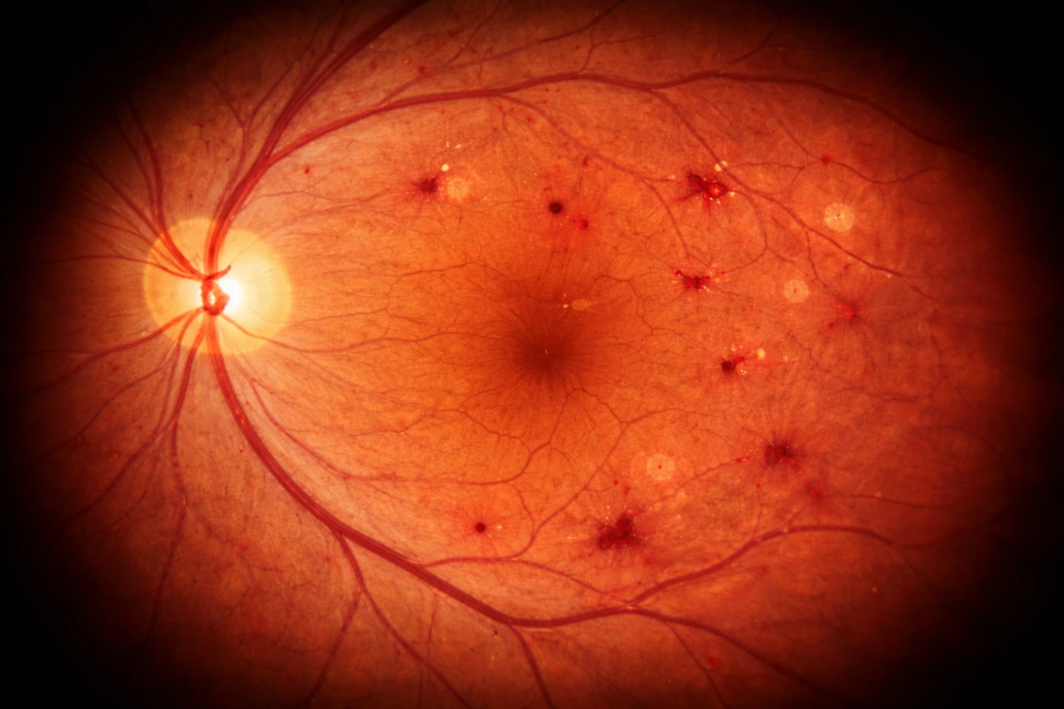
It can happen with either type 1 or type 2 diabetes, and it often starts quietly. Many people have no early symptoms because vision may still seem “fine” while changes are already happening in the retina. That is why diabetes eye screening guidelines emphasize regular dilated eye exams, since an eye doctor can spot problems before they affect daily sight.
One serious complication is diabetic macular edema, which affects the macula, the part of the retina responsible for sharp central vision. When damaged vessels leak fluid into the macula, it can swell and make reading, driving, and seeing fine detail much harder. This is one reason blurred vision with diabetes should never be brushed off, even if it comes and goes.
Why This Condition Matters
- It may begin without obvious symptoms.
- It can worsen over time and become sight threatening if it is not found and managed early.
- Early diagnosis and timely treatment lower the risk of severe outcomes.
New Findings on LRG1 in Diabetic Retinopathy
The recent buzz around new diabetic retinopathy research 2026 comes from a March 2026 research update that highlighted an underlying Science Translational Medicine paper published in 2025. The main study was done in mice, so this is preclinical evidence, but the findings are still important because they focus on the earliest stage of damage, when diabetic retinopathy may still have no obvious symptoms.
What The Study Found
Researchers used two mouse models of type 1 diabetes that mimic early retinal vascular changes seen in human disease. One was the streptozotocin, or STZ, model, and the other was the Ins2Akita model. In both models, the researchers found early induction of LRG1 after hyperglycemia developed.
Here are the findings in simple terms:
LRG1 rose early in the diabetic retinopathy mouse models. The researchers saw LRG1 activity increase at an early stage in both diabetic mouse models they studied, before the disease looked advanced.
LRG1 changes showed up before VEGF-related changes in Ins2Akita mice. In that model, LRG1 turned on first, and VEGF changes came later, which suggests LRG1 may be involved very early in the disease process.
LRG1 affected pericytes through TGF beta signaling. Pericytes are support cells that wrap around tiny retinal vessels, and the study found LRG1 changed TGF beta signaling in these cells. That shift pushed pericytes toward a tighter, more contractile state and a more fibrotic state.
Those cell changes made retinal capillaries narrower and thicker. As pericytes tightened, the retinal capillaries became more constricted. The study also reported thickening of the basement membrane, which can make the vessel wall less healthy over time.
Blood flow and oxygen delivery to the retina likely dropped. Using computational modeling, the researchers suggested these early vessel changes reduced retinal blood flow. Less flow can also mean less oxygen delivery to retinal tissue.
Blocking LRG1 helped prevent early retinal damage in the models. When the researchers removed Lrg1 or used an LRG1 function-blocking antibody, they were able to reverse or prevent early retinal vessel changes. This is why LRG1 is being discussed as a potential early therapeutic target in preclinical research.
Why This Matters For Early Vision Protection
This is the part that makes the study meaningful for patients. Current diabetic retinopathy care often focuses on detecting disease through eye exams and treating complications once they are visible or symptomatic. The LRG1 findings raise the possibility that one day doctors may be able to target an even earlier step in the disease process, before major retinal injury builds up.
It also matters because LRG1 vs VEGF diabetic retinopathy is not just a technical lab comparison. VEGF is already well known in retinal disease treatment, but this study suggests LRG1 may act earlier in diabetic retinal damage, at least in mice. If future human studies support that pattern, it could help explain why earlier intervention might protect vision better than waiting until later changes appear.
What Researchers Still Need To Prove
The next step is validation in people. Because this 2026 research on LRG1 in diabetic retinopathy new findings is based on animal models, researchers still need to confirm whether the same timing and mechanism happen in human diabetic retinopathy, whether LRG1 can reliably identify people at risk early on, and whether blocking it is safe and effective in human trials. That is an inference based on the current evidence being preclinical and UCL stating that its LRG1 targeting drug is still in further preclinical study, with human clinical trials still ahead.
So, for now, the most accurate way to read the headlines is this. LRG1 initiates early diabetic retinopathy research and scientists have found a strong early signal in mouse models that may help shape future care. It does not mean doctors are already using LRG1 in everyday screening or treatment.
Difference Between LRG1 and VEGF
When people talk about modern retinal treatment, VEGF eye therapy is usually part of the conversation. VEGF stands for vascular endothelial growth factor, a signal involved in blood vessel growth and leakage. In diabetic retinopathy and diabetic macular edema, anti VEGF eye injections are already used in real world care because they can help reduce leakage, slow disease activity, and protect vision in many patients. Current treatment options may also include corticosteroids, laser treatment, or surgery, depending on how advanced the disease is and whether there is swelling, bleeding, or traction.

What Patients Should Take Away
- VEGF is already a real treatment target in diabetic retinal disease.
- LRG1 is still in the research stage and is not yet a standard test or treatment for patients.
- The reason LRG1 is getting attention is that it may help researchers intervene earlier, not just later when symptoms or complications are easier to see.
Early Signs of Diabetic Retinopathy You Should Not Ignore
One of the hardest things about diabetic retinopathy symptoms is that there may be none at first. Early diabetic retinopathy can develop silently, which is why regular eye screening matters even when your sight seems fine. As the disease progresses, people may begin to notice changes such as blurred vision, floaters, blind spots, darker or emptier areas in vision, trouble seeing at night, faded color vision, or more general vision loss.
That means symptoms like blurry vision diabetes, eye floaters diabetes, or unexplained vision changes diabetes should not be brushed off, especially if you already have type 1 or type 2 diabetes. These symptoms do not automatically mean diabetic retinopathy, but they are worth getting checked because several diabetes related eye problems can affect vision in similar ways.
Seeing flashes or sudden new floaters and feeling unsure how urgent it is? Read what you need to know about eye flashes so you know when to call your eye doctor the same day.
Diabetic Retinopathy Stages and Complications
Doctors usually group diabetic retinopathy stages into two broad categories. The first is nonproliferative diabetic retinopathy, often shortened to NPDR. This is the earlier stage, when damaged retinal blood vessels begin to leak or close off. The second is proliferative diabetic retinopathy, or PDR. This is the more advanced stage, when new abnormal blood vessels grow in response to poor blood supply in the retina. Cleveland Clinic describes nonproliferative disease as the less severe stage and proliferative disease as the stage that causes worsening damage.
| Stage | What It Means In Simple Terms | Why It Matters |
|---|---|---|
| Mild NPDR | A few early changes, such as microaneurysms and small retinal findings | This is often the point where damage has started, even if vision still feels normal |
| Moderate NPDR | More retinal hemorrhages and signs that blood vessels are under stress | The disease is more established and needs close follow-up |
| Severe NPDR | Widespread vessel damage and poor retinal circulation | This stage carries a higher risk of progressing to sight threatening disease |
| PDR | New fragile blood vessels grow on or near the retina | These vessels can bleed or pull on the retina, raising the risk of major vision problems |
Confused by terms like low vision, impairment, and blindness when reading about complications? Start with low vision vs blindness to understand what these labels actually mean in everyday life.
How Doctors Diagnose Diabetic Retinopathy
A diabetic eye exam dilated exam is still the core way doctors screen for diabetic retinopathy. During a dilated exam, the eye doctor widens the pupil and looks at the back of the eye with special instruments. Cleveland Clinic notes that many key retinal changes do not cause symptoms until much later, but they can still be seen very early during a dilated eye exam.
That is a big reason eye screening diabetes guidelines matter. A person can feel fine and still have microaneurysms, retinal hemorrhages, abnormal blood vessels, or macular edema developing in the background. In Cleveland Clinic’s diagnostic overview, the visible early changes can include cotton wool spots, retinal bleeding, microaneurysms, neovascularization, macular edema, retinal exudates, and abnormal looking retinal blood vessels.

Common Tests Doctors May Use
Here are some of the most common parts of diabetic retinopathy diagnostics:
- Dilated retinal exam
This is the main screening and diagnostic exam because it lets the doctor directly assess the retina for early and advanced changes. - Retinal imaging
Retinal imaging can document and monitor diabetic retinopathy and diabetic macular edema over time. Cleveland Clinic notes that retinal imaging can detect both diabetes related retinopathy and diabetes related macular edema. - OCT, or optical coherence tomography
OCT is especially useful when doctors need to look for retinal swelling or monitor diabetic macular edema. StatPearls says OCT of the macula helps evaluate retinal thickening and monitor edema, while Cleveland Clinic says OCT can measure retinal thickness. - Fluorescein angiography in selected cases
When doctors need a closer look at retinal blood flow and leakage, fluorescein angiography may be used. Cleveland Clinic describes it as an imaging test that shows the blood vessels in the retina.
Why Diagnosis Often Starts Before Symptoms
One of the most important things patients should know is that doctors often find diabetic retinopathy before a person notices clear vision problems. Cleveland Clinic states this directly. Many of the key changes happen early and stay invisible to the patient at first, but they are still visible to an eye care specialist during a dilated exam. That is why screening is not just for people with blurry vision. It is also for people who think their eyes are doing fine.
Wondering if dilation is always required for a proper diabetes eye screening? Read if it’s always required to get your eyes dilated so you know when it helps most and what your doctor is looking for.
How Often Should People With Diabetes Get a Dilated Eye Exam?
A good diabetic retinopathy screening schedule depends on the type of diabetes you have, whether retinopathy is already present, and whether you are pregnant. The big takeaway is simple. Do not wait for symptoms. How often should people with diabetes get a dilated eye exam is one of the most important prevention questions because early retinal changes can be found before vision starts to blur. ADA 2026 guidance and the AAO both still center care around a comprehensive dilated eye exam, with shorter follow-up if retinopathy is progressing or other risk factors are raising concern.

If you have type 1 diabetes
Plan for your first comprehensive dilated eye exam about 5 years after diagnosis. That timing reflects the fact that diabetic retinopathy usually takes time to develop after hyperglycemia begins.
If you have type 2 diabetes
Get a comprehensive dilated eye exam at the time of diagnosis. Type 2 diabetes can be present for years before it is officially diagnosed, so retinal damage may already be there when someone first learns they have diabetes.
For ongoing follow-up
Most adults with diabetes should continue with yearly dilated eye exams. If one or more annual exams show no retinopathy and blood sugar markers are staying in goal range, ADA 2026 says screening every 1 to 2 years may be considered for some lower risk patients. On the other hand, follow-up needs to be more frequent if retinopathy is getting worse or if risk factors such as not meeting glycemic goals are present.
If you are pregnant and already have type 1 or type 2 diabetes
Eye care should start before pregnancy if possible, or early in the first trimester if you are already pregnant. Close follow-up during pregnancy is important because diabetic retinopathy can worsen during this time, especially if retinopathy is already present. ADA and AAO guidance also note that follow-up continues during pregnancy and may extend into the postpartum period based on how much retinopathy is present.
If you have gestational diabetes alone
This is different from preexisting diabetes. ADA 2026 and AAO guidance say that people who develop gestational diabetes mellitus alone generally do not need routine retinopathy eye exams during pregnancy because they do not appear to be at increased risk for diabetic retinopathy during that pregnancy.
If you are unsure when to see an eye doctor for diabetic retinopathy, the safest move is to ask your diabetes clinician or eye doctor to set your next exam before you leave the visit.
When to See an Eye Doctor
Not every vision change is an emergency, but some changes should never be brushed off. The easiest way to think about it is this. Gradual changes usually mean you should book an eye exam soon. Sudden changes can mean you need urgent or emergency care. That is especially important for those looking up early signs of diabetic retinopathy and when to seek care because diabetic eye disease is not the only possible cause of vision symptoms, and some retinal problems need immediate treatment.

Book An Eye Exam Soon If You Notice
If your vision changes gradually, do not wait months to see whether it settles down. Cleveland Clinic advises calling or seeing your provider if you notice vision loss or reduced clarity, blurred or distorted vision, or areas of sight that look dim, faded, or different from before.
- Gradual blurry or distorted vision that keeps coming back or slowly worsens. This fits the kind of change Cleveland Clinic says should prompt a provider visit.
- New dim, faded, or patchy areas in your sight even if the change is subtle at first.
- Worsening trouble seeing clearly, especially if reading, driving, or recognizing detail suddenly feels harder than usual.
This is where people often search for phrases like sudden blurry vision in diabetes when to see an eye doctor, but the timing matters. If the blur is gradual, book a prompt exam. If it is dramatic and sudden, treat it as urgent.
Not sure whether to book with an optometrist or go straight to an ophthalmologist when symptoms feel serious? Identify who should you see between ophthalmologist and optometrist to choose the right next step.
What to Do If You Notice Vision Changes With Diabetes
If your vision starts looking spotty, hazy, dim, or blurry, do not assume it will “just clear up.” Diabetic retinopathy can be present with no obvious symptoms at first, and many of the early changes can still be seen during a dilated eye exam.
Take the changes seriously
Gradual vision changes like blur, distortion, or areas that look dim or faded are worth acting on, even if they come and go. Those symptoms can show up when the retina is under stress from diabetes, and waiting can mean missing the best window to catch problems early.
Book a dilated eye exam soon
A dilated exam gives your eye doctor a clear view of the retina, where diabetic retinopathy and diabetes-related swelling can start. Cleveland Clinic notes that key retinal changes may be visible early on to an eye care specialist, even before symptoms become obvious.
Keep the rest of your diabetes care on track
Eye health is closely tied to whole-body control. The ADA Standards of Care in Diabetes 2026 recommends working toward glycemic goals, as well as blood pressure and lipid goals, to reduce risk or slow progression of diabetic retinopathy.
Follow through on recommended testing
If your eye doctor suggests retinal imaging, do not put it off since it can document retinopathy changes and help monitor progression. OCT is especially useful when swelling is a concern because it can measure retinal thickness and help evaluate diabetes-related macular edema.
Keep research updates in perspective
Research like the recent LRG1 findings is promising, but it is not a substitute for care. In mouse models, researchers found LRG1 may play a role very early in diabetic retinal damage, and blocking it prevented early damage in those models, which is why it is being discussed as a potential pre-emptive therapeutic target.
The practical takeaway is still the same: if your vision changes with diabetes, your next step is getting your eyes checked rather than trying to self-diagnose.
Did your vision symptoms start after a new prescription or dosage change? Review the medications that cause vision problems so you know what to mention at your appointment.
Current Treatment Options Available Today
Treatment depends on what the retina looks like in real life, not just on the diagnosis label alone. Doctors consider the stage of retinopathy, whether there is diabetic macular edema, whether blood vessels are leaking or growing abnormally, and whether complications such as vitreous hemorrhage or retinal detachment are present.
1. Anti VEGF Injections

Anti VEGF injections are one of the best known modern treatments for diabetic retinal disease, especially when swelling or abnormal vessel activity is involved. AAO notes that anti VEGF treatment improves vision in about one third of patients and at least stabilizes vision in most others. AAO also notes that laser may be used to reduce retinal swelling, but anti VEGF therapy is a central part of current treatment conversations for diabetic retinopathy and diabetic macular edema.
2. Corticosteroids in Selected Cases

Some patients may be treated with corticosteroids, especially when inflammation is part of the picture or when another approach is not the best fit. Cleveland Clinic lists corticosteroids among medication options for diabetic retinopathy, and AAO reviews note that intravitreal corticosteroid therapy is used for selected patients with diabetic macular edema.
3. Laser Treatment

Laser treatment still has an important role in care. According to Cleveland Clinic and AAO, laser photocoagulation can be used to prevent blood vessel growth, shrink abnormal vessels, or seal leaking vessels and reduce swelling in the retina. In plain language, laser is often used when the goal is to control leakage or reduce the effect of abnormal retinal blood vessels.
4. Vitrectomy or Other Retinal Surgery

When disease becomes more advanced, diabetic retinopathy surgery may be necessary. Cleveland Clinic says a vitrectomy may be recommended if the retina is detached, if there is vitreous hemorrhage that is not clear, or if there is certain other retina damage. This is usually the kind of treatment doctors consider when bleeding, traction, or detachment is threatening vision more seriously.
The best option depends on the stage of disease, the amount of swelling, whether bleeding is present, and whether serious retinal complications have developed. That is why treatment decisions are based on your eye exam and imaging, not just on symptoms alone.
What the LRG1 Research Could Change Next
The March 2026 update found that LRG1 appears very early in diabetic retinal damage in mouse models, before VEGF related changes became prominent in one of the models. That is why researchers are paying attention to it as a possible earlier point of intervention. But the evidence is still preclinical, which means it is not yet a routine test, treatment, or early warning tool for patients.
LRG1, Diabetic Retinopathy, and the Importance of Acting Early
The recent attention around LRG1 is worth watching because it points to a more detailed understanding of how early diabetic retinopathy may begin.That makes the research promising, but it does not mean LRG1 is already a routine screening marker or a standard treatment for patients today.
Keep up with regular dilated eye exams, because diabetic retinopathy can start before obvious symptoms appear. Do not ignore new blur, distortion, faded areas, floaters, or other vision changes diabetes can cause. And if you ever have sudden severe vision loss or severe eye pain, treat that as urgent and get immediate care.
The science is moving forward, and researchers may be getting closer to earlier ways to detect or treat diabetic eye disease. But the best way to protect your sight today is still simple and proven: stay on top of diabetes care, follow your eye exam schedule, and act quickly when your vision changes.
Need to double-check the numbers on your prescription before you shop for contacts online? Learn how to read contact lens prescriptions (OD vs. OS) so you enter the correct details for each eye.
Has your vision changed, or are you due for an updated prescription? Start with a comprehensive eye exam so you know exactly what your eyes need. Then, if your prescription is current and you are ready to shop, browse our contact lens selection to find options that fit your prescription, comfort preferences, and everyday routine.
FAQs about LRG1 in Diabetic Retinopathy
What is LRG1 and how is it linked to diabetic retinopathy?
LRG1 is a protein that researchers found to be involved very early in diabetic retinal blood vessel dysfunction in mouse models. In the 2025 Science Translational Medicine paper highlighted again in March 2026, LRG1 rose early, contributed to capillary narrowing and reduced oxygen delivery, and blocking it helped prevent early damage in those models.
Is LRG1 a proven early biomarker for diabetic retinopathy in humans?
No, not at this point. The current evidence behind the 2026 headlines comes from preclinical animal research, not from a validated human screening test. That means LRG1 is promising, but it is not yet a proven biomarker doctors use in routine patient care.
How is LRG1 different from VEGF in diabetic eye disease?
VEGF is already part of current retinal treatment because anti VEGF medicines can reduce leaking abnormal blood vessels and help manage diabetic macular edema and some diabetic retinopathy cases. LRG1 is different because the mouse study suggests it may act earlier in the disease process, before VEGF related changes become more prominent.
Can diabetic retinopathy start before symptoms appear?
Yes. Early diabetic retinopathy often has no obvious symptoms, which is why regular eye screening matters so much. Both Cleveland Clinic and the ADA note that important retinal damage may be present before a person notices blur, floaters, or other vision changes.
What are the earliest warning signs of diabetic retinopathy?
The earliest stage may not cause symptoms, but common warning signs as disease progresses include blurred or distorted vision, faded colors, trouble seeing at night, floaters or streaks, blind spots, and vision loss. Diabetes related macular edema can also cause blur, dark spots, glare problems, and lines that look bent or curved.
How often should people with diabetes get a dilated eye exam?
For adults with type 2 diabetes, the first eye exam is recommended at diagnosis. For people with type 1 diabetes, the first exam is generally recommended within about five years of diagnosis. After that, most people need at least yearly follow up, though some lower risk patients may be seen every one to two years based on findings and overall control.
When is blurry vision from diabetes an emergency?
Gradual blurry or distorted vision should prompt an eye exam soon. But sudden severe vision loss or severe eye pain should be treated as an emergency. Cleveland Clinic specifically advises going to the nearest hospital or emergency room for sudden severe vision changes like those.
Do new floaters and flashes with diabetes need urgent care?
Yes, especially if they start suddenly. The AAO warns that sudden floaters and flashes can be symptoms of a torn or detached retina, and Cleveland Clinic notes that a sudden increase in floaters can be a sign of retinal tear or detachment that needs immediate treatment.
Can diabetic retinopathy be treated or reversed?
Diabetic retinopathy is treatable, but it is not considered curable. Cleveland Clinic says some symptoms and retinal changes can be treated, but certain damage is not reversible once it becomes severe enough. That is why AAO stresses getting treatment as early as possible to help prevent vision loss.
What should I do if my vision becomes hazy, patchy, or distorted?
Do not wait to see if it goes away on its own. Book a dilated eye exam promptly, follow through with any retinal imaging your doctor recommends, and stay on track with blood sugar, blood pressure, and cholesterol goals. The ADA notes that eye exams, early treatment, and managing glucose, blood pressure, and cholesterol are key steps in preventing vision loss from diabetes related eye disease.
Disclaimer: This content is for general informational purposes only and is not a substitute for professional advice, diagnosis, or treatment.
References
Leucine-rich α-2-glycoprotein 1 initiates the onset of diabetic retinopathy in mice. PubMed / National Library of Medicine. https://pubmed.ncbi.nlm.nih.gov/41124286/. Date Accessed: March 11, 2026.
Retinopathy, Neuropathy, and Foot Care: Standards of Care in Diabetes—2026. Diabetes Care / American Diabetes Association. https://diabetesjournals.org/care/article/49/Supplement_1/S261/163919/12-Retinopathy-Neuropathy-and-Foot-Care-Standards.Date Accessed: March 11, 2026.
Diabetic Retinopathy. National Eye Institute (NIH). https://www.nei.nih.gov/eye-health-information/eye-conditions-and-diseases/diabetic-retinopathyDate Accessed: March 11, 2026.
Vision Loss and Diabetes. Centers for Disease Control and Prevention. https://www.cdc.gov/diabetes/diabetes-complications/diabetes-and-vision-loss.html.Date Accessed: March 11, 2026.
VEHSS Modeled Estimates: Prevalence of Diabetic Retinopathy (DR). Vision and Eye Health Surveillance System / Centers for Disease Control and Prevention. https://www.cdc.gov/vision-health-data/prevalence-estimates/dr-prevalence.html.Date Accessed: March 11, 2026.
Diabetic Retinopathy: Causes, Symptoms, Treatment. American Academy of Ophthalmology. https://www.aao.org/eye-health/diseases/what-is-diabetic-retinopathy.Date Accessed: March 11, 2026.
Diabetic Retinopathy PPP 2024. American Academy of Ophthalmology. https://www.aao.org/education/preferred-practice-pattern/diabetic-retinopathy-ppp.Date Accessed: March 11, 2026.
Injections to Treat Eye Conditions. National Eye Institute (NIH). https://www.nei.nih.gov/eye-health-information/eye-conditions-and-diseases/diabetic-retinopathy/injections-treat-eye-conditions.Date Accessed: March 11, 2026.
Diabetes-Related Retinopathy: Symptoms, Stages & Treatment. Cleveland Clinic. https://my.clevelandclinic.org/health/diseases/8591-diabetic-retinopathy.Date Accessed: March 11, 2026.
What Are Floaters and Flashes? American Academy of Ophthalmology. https://www.aao.org/eye-health/diseases/what-are-floaters-flashes.Date Accessed: March 11, 2026.